CASE STUDY
Custom Aneurysm Treatment that Saves Lives
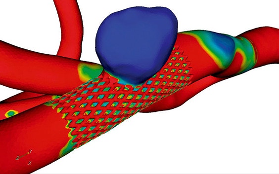
At the University of Limerick, Ireland, a Finite Element Analysis Rupture Index (FEARI) was created to help the surgical decision-making process by expressing the rupture potential of an aneurysm. Together with 3D reconstructions, this can play a key role in improving the treatment of abdominal aortic aneurysms (AAAs).
The challenge
Assess the role of FEARI and 3D reconstruction in treating AAAs
Cardiovascular disease is a leading cause of premature death. It kills around 900,000 people per year in the United States alone, with ruptured aneurysms forming a significant part of these deaths.
Researchers at the University of Limerick, Ireland, created a novel index called the Finite Element Analysis Rupture Index (FEARI) to help the surgical decision-making process by expressing the rupture potential of an aneurysm.
As during the surgical intervention, the correct sizing of the medical device is of utmost importance to avoid complications, the two aims of this study were first to examine FEARI’s effectiveness and second to show how 3D reconstructions created in Materialise Mimics can be a useful surgical guidance tool.

The solution
3D reconstructions from CT data using Mimics
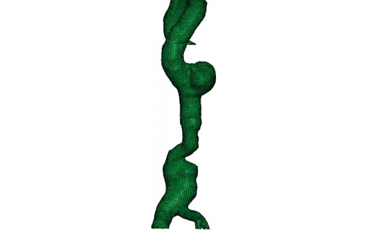
For this study, CT data was collected from ten patients suffering from abdominal aortic aneurysms (AAA) and imported into Mimics. Within only a few steps, a 3D reconstruction was generated.
First, a thresholding technique was applied to the data to highlight the areas of interest. Second, these areas were segmented with different colors being assigned to specific features, for example, the diseased aorta. Finally, with the segmentation done, a 3D model was quickly and easily created.
“3D reconstruction of AAAs using Mimics is a powerful and useful tool. It allows the surgeon to obtain measurements useful for the sizing of stent grafts and also allows for the visualization of the aneurysm prior to surgery.”
— Barry Doyle and Timothy McGloughlin, University of Limerick
In the case of the aorta, reconstruction times ranged from two minutes for basic models where minor details were ignored to one hour for a model where all details were included, such as intraluminal thrombus and surface indentations. As CT scans are taken a considerable time prior to the actual operation, and thanks to the possibility to quickly create 3D reconstructions, surgeons can easily afford to examine a 3D model without sacrificing the health status of the patient.
The FEARI involves an equation based on a simple engineering definition of material failure; that is, failure will occur when the stress acting on the material exceeds the strength of the material.
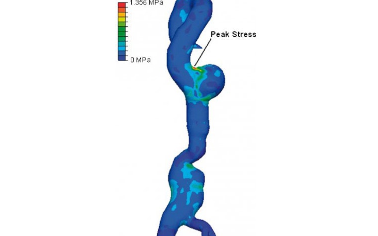

As such, the value of FEARI is calculated by dividing the finite element analysis (FEA) wall stress by the experimental wall strength. The result is a value between 0 and 1, with 0 indicating a low rupture potential, and values close to 1 indicating a very high rupture potential.
While the experimental wall strength values were obtained from previous research, the FEA wall stress values were obtained using the ten 3D reconstructions generated in Mimics. The 3D reconstructions were imported into an FEA program for stress analysis, the resulting values being used in the FEARI calculation.
The result
Improved surgical planning and outcome
After analyzing the values obtained during this study, it was concluded that FEARI could indeed serve as a useful tool for surgical decision-making when used in conjunction with current procedures.
Barry Doyle and Timothy McGloughlin from the University of Limerick, Ireland, explain: “3D reconstruction of AAAs using Mimics is a powerful and useful tool. It allows the surgeon to obtain measurements useful for the sizing of stent grafts and also allows for the visualization of the aneurysm prior to surgery. Reconstructions are also necessary for further use with FEA, which has been shown to be a good method of determining wall stress in the diseased aorta. Because of this, 3D reconstruction could aid surgeons in improving the treatment of AAAs.”
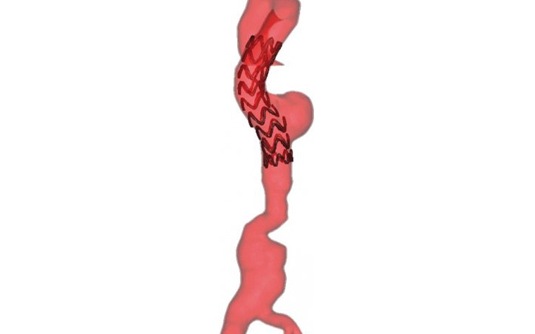

When the decision is made to operate, the preferred surgical intervention is endovascular aneurysm repair (EVAR). Many benefits were found when EVARs were planned using Mimics and 3D reconstructions.
The sophisticated measurement tools in Mimics provide accurate dimensions of complex geometries, allowing for accurate measurements and greater insight into the morphology of the diseased aorta. This is important as surgeons are tackling more challenging anatomy than ever with EVAR. The valuable information from the 3D reconstructions ensures that the surgeons do not encounter any unforeseen problems during surgery; this, in turn, increases confidence in the procedure.
Additionally, the precise measurements that can be determined using Mimics aid in the choice of the most appropriate stent graft. Furthermore, it means that new medical devices can also be conceived and designed for a better fit for patients. Finally, post-operative 3D reconstructions can also help the surgeon observe the outcome of the EVAR procedure at regular intervals, allowing for quick identification and treatment of complications.
In conclusion, Mimics’ powerful 3D reconstruction tool has proven highly useful. It serves a role in calculating FEARI, which has the potential to aid in the surgical decision-making process. Furthermore, 3D reconstruction is performant and could not only help to standardize the measurement and size stent grafts, but also support surgeons in improving AAA treatment.
L-102654-01
Share on:
This case study in a few words
Healthcare
Materialise Mimics
Powerful 3D visualization
Accurate measurement of complex geometries
