EXPERT INSIGHT
7 Ways 3D Planning Can Support Your LAAO Procedure

Mimics Enlight’s LAAO planner joins the TMVR workflow for creating clear pre-procedural plans by using 3D planning. Find out what the LAAO planner can do for you.
Not all 3D is the same
The medical sector, and particularly the cardiovascular field, is tapping into innovative technology to deliver personalized and predictive planning to support clinicians in providing better care for their patients. However, not all planning tools are equal or even using the same technology.
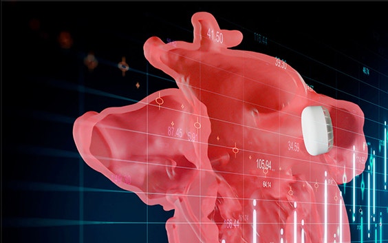
Mimics Enlight recently released the new left atrial appendage occlusion (LAAO) planner, joining their TMVR workflow. The two workflows assist you in creating clear pre-procedural plans with the use of 3D planning. This allows clinicians to plan and truly measure in 3D as opposed to 2D planning with volume rendering for visualization purposes only. Professor Nicholas Van Mieghem, the Medical Director of Interventional Cardiology at Erasmus UMC, explains, “The anatomical appreciation is crucially different between conventional CT and with 3D planning. We added so much more granularity to our understanding using the 3D planning."
New technology is supporting treatments in ways that help mitigate the risk for patients and improve efficiency. Read on to find out what the new LAAO planner can do for you using this technology.
1. Enhanced 3D visualization
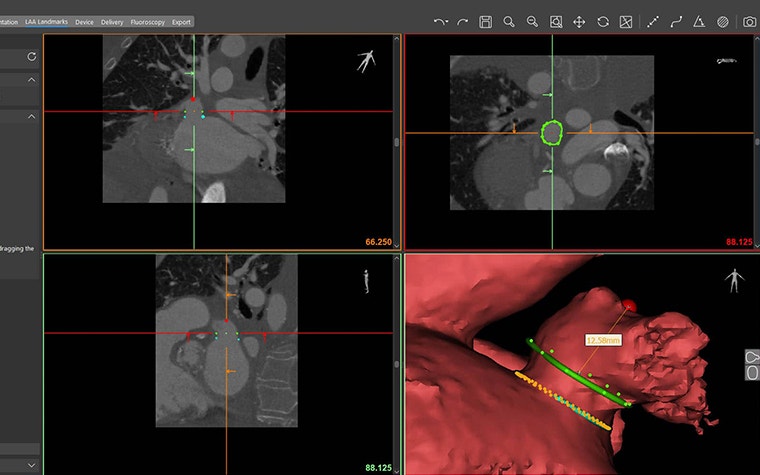
Instantly gain an accurate understanding of the LAA anatomy in 3D. You’ll be able to easily assess the relationship between the LAA and surrounding structures with comprehensive visualization and measurements directly in 3D.
"The anatomical appreciation is crucially different between conventional CT and with 3D planning," says Prof. Nicolas Van Mieghem, Interventional Cardiology at Erasmus University Medical Center. "We added so much more granularity to our understanding using the 3D planning."
2. Landing zone assessment in 3D
You’ll be able to simply indicate the LAA ostium and landing zone on CT or directly on the interactive 3D model and automatically receive accurate area measurements. Visualize and adapt the area in 3D with automatic measurements updates so you can properly assess what you’re going into with the procedure.
3. Automatic appendage depth measurement
Assessing the appendage depth with the intuitive automatic depth measurement tool will be an easy process. Accurately measure the appendage depth by planning in 3D and receiving real-time, updated depth measurements as you adjust your planning.


4. Virtual implantation
Selecting the correct device and illuminating challenging delivery pathways is straightforward by using enhanced 3D visualization. Indicative simulation of device delivery will provide you with an understanding of device feasibility and implant fit.
5. Online case sharing
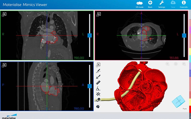
Connect with your heart team on procedural plans with one-click report generation and virtual case sharing with Mimics Viewer. The online platform is optimized for desktop and mobile users and allows the receiver to view your 3D patient models, planning, and device designs at their convenience via a user-friendly interface.


6. Virtual reality
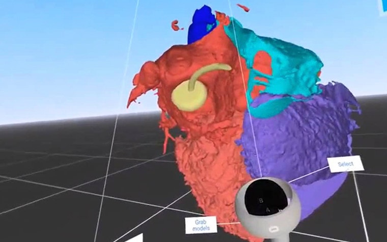
Enhance your virtual experience with Mimics Viewer by exploring your case in VR. You’ll be able to truly appreciate the three-dimensional structure.


7. 3D printing
Physically assess implantation and catheter feasibility for complex cases by using an accurate 3D-printed copy of your patient’s anatomy. This is made possible by easily exporting your planning for 3D printing at the point of care or by using Materialise’s HeartPrint service.
L-101801-01
Share on:
You might also like
Never miss a story like this. Get curated content delivered straight to your inbox.
