EXPERT INSIGHT
6 Questions to Ask Before Starting with 3D Printing in Your Hospital Part 2
As we saw in the first part of this series, there are several considerations to take into account before implementing 3D printing in your hospital. In this post, we will discuss in further detail how you can create an accurate file for 3D printing. We’ve already covered image processing, so here are three more potential problems that you might face, along with the strategies to overcome them.
What software tools will you need throughout the workflow?
3D modeling
Segmenting the medical images is just the beginning. The most laborious part of the 3D modeling process is often the further preparation and augmentation of your segmented anatomy. This can include features such as cleaning and smoothing to remove artifacts, adding connectors to hold anatomies in the proper position, adding thickness to represent vessel walls, cutting the model to achieve optimal visualization, as well as indicating color or multiple materials.
It is essential to take all these considerations into account, and the way you prepare your 3D model will determine how useful the print will be clinically, how much time and material will be required to build the part, and the feasibility of printing and cleaning the eventual model.
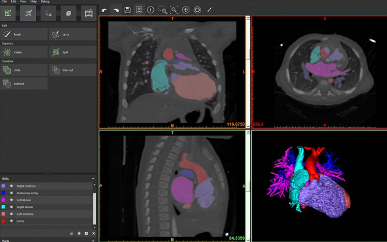

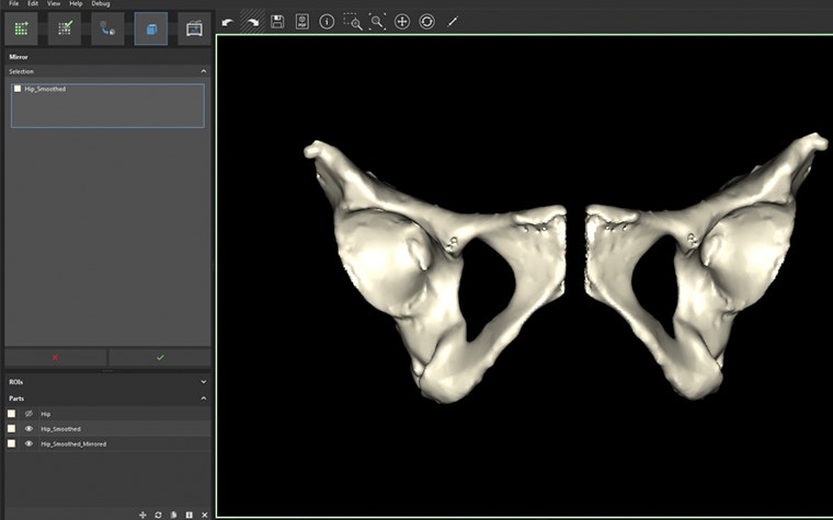
For example, if a surgeon requests a model to plan a trauma reparation, they might want to understand the optimal outcome of the restoration. For this, it would be helpful to provide a second model of the patient’s mirrored anatomy to understand what the optimal outcome would look like. For a complex heart procedure, the surgeon will want to understand the intricacies of the intra-cardiac anatomy or the landmarks for a specific valve or artery. This will require you to prepare the heart model with windows or to cut along a split line to achieve this visualization and separate components to identify another color in the model. Each 3D printing request will have very different 3D modeling requirements.
Verification and labeling
After completing your segmentation and modeling work, you might want to get your part printed as soon as possible. However, it is easier than you might think to make mistakes during the previous steps. Prior to printing, the accuracy of your final file should be verified against the original DICOM imaging. Did you over-smooth and remove a key feature from the model? Did you cut away a structure that would be an important landmark for the surgeon? From previous experience, a solution that allows the user to overlay the STL surfaces back on the DICOM data is the most efficient. This will allow you to verify accuracy (and convince others of it) as well as make subtle adjustments or refinements to the model.
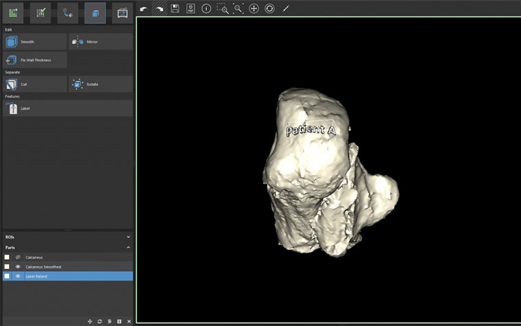

Ensuring the traceability of your 3D prints will also reduce the opportunity for making errors with your 3D printing program. As you scale your operation and build greater volumes of models, it is critical to understand what part is coming off the printer and what case it belongs to. To reduce the chance of mixing models or providing the wrong model to a surgeon, each anatomical model should be pre-labeled in the software prior to printing. Use a requisition number that will trace back to the medical records to ensure the traceability of your 3D models.
You will also need to use labels if you create mirror images or want to clearly indicate what side of the patient the model was derived from. This will reduce the chances of the surgeon getting confused and prevent them from operating on the wrong side of the body.

3D file fixing
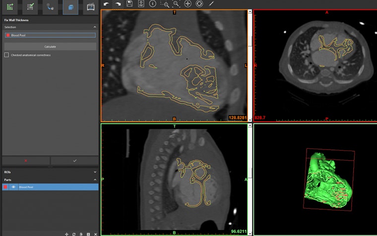
The STL file format is the universal digital 3D modeling format for 3D printing. This is the file that will be fed to the 3D printer to slice and build the part. However, not all STL files are created equal. The number and quality of the triangle facets will determine the eventual quality of your printed part. You may also find very thin walls in the model that fall under the minimum resolution of your printer or that will be very brittle and tear-sensitive. It is imperative to have a robust STL diagnostic and fixing tool to ensure a successful and quality build as well as making sure that all walls are printed.

Check out our third and final blog post, in which you will find the answer to any questions you may have about 3D printers, communication, personnel, and training! Or watch the webinar recording on implementing 3D printing in your hospital to learn more.
This blog is based on an article first published on auntminnie.com
L-102879-01
Share on:
You might also like
Never miss a story like this. Get curated content delivered straight to your inbox.
