EXPERT INSIGHT
5 Quick Tips to Improve Your Medical Image Segmentation Process

Designing a personalized device, performing an anatomical study, creating a virtual model for 3D printing, performing an FEA study. None of these applications would be possible without an accurate segmentation of the anatomy of interest from medical imaging data. Indeed, segmentation can rightfully be called the basis for all of these! Today, we’ll share some of the most important aspects to take into account for optimizing your segmentation process, in terms of accuracy as well as speed.
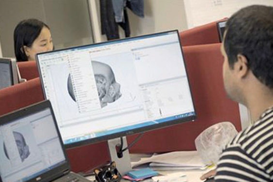
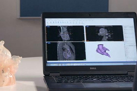
1. Make sure the input data is of the highest possible quality
Choose the appropriate modality — usually CT or MRI — to obtain what you need to know from a scan. Stick to a consistent protocol to avoid variation. It is worth your while to invest some time into developing a high-quality imaging protocol together with the Radiology department and using sources from scientific literature. If you are working with multiple centers, make sure that the same protocol can be applied everywhere by, for example, fine tuning the protocol so that it works on machines from different manufacturers. Without the necessary slice thickness, slice increment, or the right reconstruction algorithms, it will never be possible to reach a sufficient level of segmentation quality for surgeons’, patients’, or your own needs. Questions on how to find the perfect scanning protocol or image acquisition method? Don’t hesitate to get in touch with your Materialise representative!
2. Organize your workflow
When it comes to segmentation quality, one of the largest sources of variation is the different approaches that team members take to a segmentation task. Materialise Mimics offers several tools that can help you with this. ‘MyTab’ allows you to create your own custom toolbar. So if you prefer to use your favorite tools in a certain order or would like other team members to use them in that order as well, you can create your own custom workflow. If you want to increase speed and take even more variables out of your segmentation workflow, you could consider using the extensive automation options offered in the Scripting module.
Practically all steps at the beginning — such as importing DICOM images, anonymizing, creating initial masks — and the end — like wrapping, smoothing, exporting to STL — can be automated, saving you precious time and increasing consistency as the script can be made in such a way that it always uses the same settings.
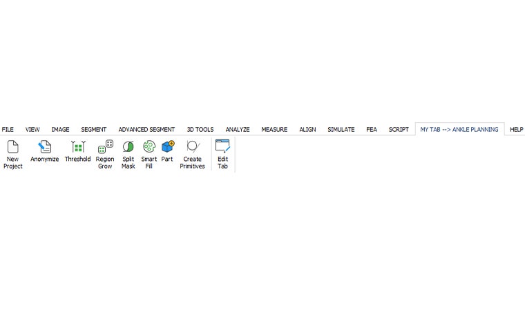

3. Use the latest, most powerful tools
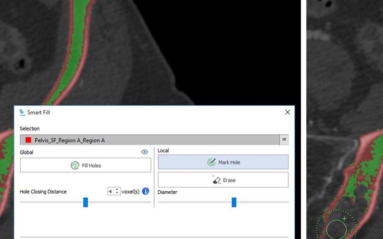
Split Mask and Smart Fill are some of our favorite tools for segmenting bones from CT. If you are dealing with scans of osteoarthritic patients and Region Growing is not enough to separate the two bones, Split Mask is ideal for separating the two bones in a joint in just a few clicks. Then, if you want to obtain filled bone masks, use the Smart Fill tool to fill up the bone much faster than with previous tools. Combined, these two tools can save you many minutes per scan. If you are working on 4D CT data of the heart, you will be happy to know that you can import 4D CT scans into Mimics and easily copy-paste masks between phases to immediately obtain an initial segmentation of that next frame. Or, use the 4D CT Heart tool to segment all phases of the cardiac cycle in one go.

4. Get proper training
We cannot stress enough how much time you might be able to save on your projects with the proper training for the type of work that you do. Mimics is a toolbox that can sometimes be daunting to get started with. Look here to find training courses near you, or order an on-site session for customized training.
5. Stay tuned for insights from Materialise
On this site, via email, and at events throughout the world. We release new versions of Mimics every year, and each release always contains improvements in segmentation. Furthermore, we also regularly offer customers the opportunity to try out new tools before they are officially released.
L-102647-02
Partageons :
Vous aimerez peut-être aussi
Ne ratez jamais une histoire comme celle-ci. Recevez-les dans votre boîte de réception une fois par mois.
